This program was part of the 2026 edition (June 9–11, 2026). Discover the 2027 edition
Pre-congress course: Imaging, theory (40 places) and practice (9 places)
-
Date
June 9–11, 2026
-
Duration
3 Intensive Days
ABOUT THE COURSE
Comprehensive Training
Surgery for deep endometriosis has evolved significantly in recent years, and one of the main pillars of this evolution is improved preoperative diagnosis and staging through imaging. Well-performed mapping using ultrasound and/or magnetic resonance imaging allows more precise characterization of the disease, anticipation of technical complexity, estimation of risks, guidance on the need for a multidisciplinary team and support for clearer, more realistic discussion with the patient about the benefits, limitations and potential complications of the proposed treatment.
In this context, the imaging specialist plays a central role in the endometriosis care pathway: contributing to initial assessment, severity stratification, objective communication with the surgeon and follow-up after medical or surgical treatment. At the same time, it is important to recognize that examinations have pitfalls and limitations, and that the quality of the result depends on technique, standardization, report language and integration with clinical findings.
This course was developed to present the practical foundations of endometriosis mapping using ultrasound and magnetic resonance imaging, integrating theoretical classes with practical demonstrations, case discussions and training in structured reporting oriented toward treatment planning.
ACTIVITIES
Course Coordination
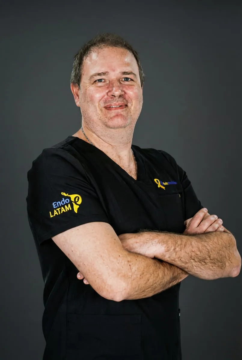
- Dr. Carlos Henrique Trippia
Guest faculty
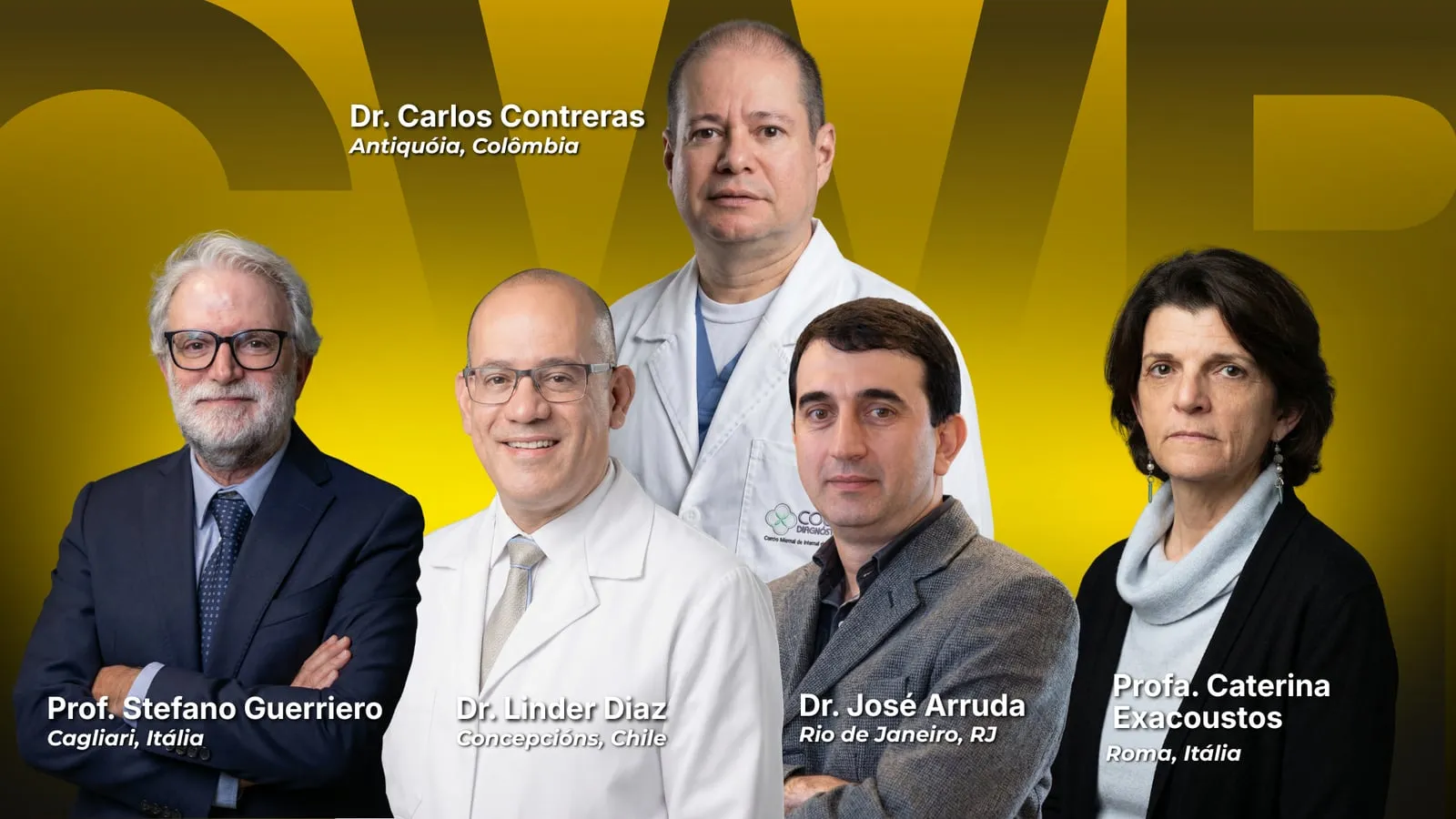
National and International Guests
OBJECTIVES
What you will learn
By the end of the course, participants will be able to:
- Diagnose adenomyosis and perform the differential diagnosis with leiomyomas, recognizing key signs and situations of diagnostic overlap.
- Assess and characterize ovarian endometriomas, discussing relevant differential diagnoses and findings that affect management.
- Identify deep endometriosis at different sites and describe signs of involvement and extent using reproducible language.
- Recognize pitfalls, limitations and common sources of false-negative/false-positive findings, qualifying the examination conclusion with clinical responsibility.
- Prepare a structured, objective report focused on investigating endometriosis, with essential items for communication with the care team and surgical planning.
- 3 DAYS OF IMMERSION
- 6 FACULTY
IMMERSION
Schedule
-
08:00 – 08:10
Welcome, Introduction and Course Overview
Dr. Carlos Henrique Trippia
-
08:10 – 09:10
Normal pelvic anatomy on imaging
-
08:10 – 08:30
Pelvic anatomy of the anterior and middle compartments
- Speaker to be confirmed
-
08:30 – 08:50
Pelvic anatomy of the posterior and lateral compartments
- Speaker to be confirmed
-
08:50 – 09:05
Neural anatomy on ultrasound
- Speaker to be confirmed
-
09:10 – 09:30
Adenomyosis and its clinical presentations
Profa. Caterina Exacoustos – Roma, Itália
-
09:30 – 09:50
Diagnosis of Müllerian anomalies by ultrasound and/or pelvic MRI
- Speaker to be confirmed
-
09:50 – 10:10
Uterine fibroids: how to help the surgeon with surgical planning
- Speaker to be confirmed
-
10:15 – 10:45
Coffee break
-
10:45 – 11:00
Adnexal endometriosis and atypical patterns of ovarian endometriomas
Profa. Caterina Exacoustos – Roma, Itália
-
11:00 – 11:15
Tubal assessment by imaging methods before and after surgery
- Speaker to be confirmed
-
11:20 – 12:00
Endometriosis mapping: how I do it
-
11:20 – 11:40
By ultrasound
- Speaker to be confirmed
-
11:40 – 12:00
By MRI
- Speaker to be confirmed
-
12:00 – 12:30
Interactive roundtable (Questions and answers)
-
12:30 – 13:45
Lunch
-
13:45 – 14:00
Endometriosis and systematic approach: IDEA consensus step 1 and 2
Prof. Stefano Guerriero – Cagliari, Itália
-
14:00 – 14:15
Endometriosis and systematic approach: IDEA consensus step 3 and 4
Profa. Caterina Exacoustos – Roma, Itália
-
14:15 – 14:30
Discussion
-
14:30 – 15:30
Classes, clinical cases and ultrasound examination videos
- In-person session with all faculty
- Coordination: Dr. Linder Diaz
-
15:30 – 15:50
Coffee break
-
15:50 – 16:10
Superficial endometriosis on ultrasound: diagnostic criteria and technical limitations
Prof. Stefano Guerriero
-
16:10 – 18:30
Classes, clinical cases and ultrasound examination videos
-
16:10 – 17:10
In-person session with all faculty
- Coordination: Dr. José Arruda
-
17:10 – 18:30
In-person session with all faculty
- Coordination: Prof. Stefano Guerriero & Profa. Caterina Exacoustos
-
18:30
End of the course for the Practical Course
-
08:00 – 09:00
Endometriosis findings in:
-
08:00 – 08:20
Anterior compartment
- Speaker to be confirmed
-
08:20 – 08:40
Posterior compartment
- Speaker to be confirmed
-
08:40 – 09:00
Lateral compartment
- Speaker to be confirmed
-
09:20 – 09:40
ENZIAN classification and its application in imaging examinations
- Speaker to be confirmed
-
09:40 – 09:45
Practical demonstration of ENZIAN application
- Speaker to be confirmed
-
09:45 – 10:00
Markers of deep endometriosis (indirect signs of endometriosis)
- Speaker to be confirmed
-
10:00 – 10:20
Coffee break
-
10:20 – 10:40
Unusual sites of endometriosis (abdominal wall, right iliac fossa, diaphragm and chest, etc.)
- Speaker to be confirmed
-
10:40 – 11:00
Postoperative follow-up imaging: Tips and tricks to differentiate postsurgical fibrosis, persistence or recurrence of the disease.
- Speaker to be confirmed
-
11:00 – 12:00
Interactive roundtable: Questions and answers
-
12:00 – 13:30
Lunch
-
13:30 – 13:50
Pelvic neurosonography: the state of the art
Dr. José Arruda
-
13:50 – 14:10
Strategies to increase diagnostic accuracy in US/MRI mapping (systematic protocol, adjunct techniques, structured report, use of AI).
-
14:10 – 14:30
Discussion
-
14:30 – 15:10
Classes, clinical cases and ultrasound examination videos correlated with MRI
- In-person session with all faculty
- Coordination: Dr. Carlos Henrique Trippia
-
15:10 – 15:30
Coffee break
-
15:30 – 16:30
Classes, clinical cases and ultrasound examination videos correlated with MRI
- In-person session with all faculty
- Coordination: Dr. Carlos Contreras & Dr. Carlos Trippia
-
16:30
End of the course for the Theoretical Course
-
17:15
Social gathering
-
08:00 – 17:30
Supervised hands-on at Clínica IR Diagnóstico
Dr. Carlos Henrique Trippia
Profa. Caterina Exacoustos
Dra. Lilian Aparecida Affonso Fazzion
Dr. José Arruda
Dr. Linder Díaz
Prof. Stefano Guerriero
Dr. Carlos Contreras
-
08:00 – 12:00
Imaging examinations performed under faculty supervision
-
12:00 – 13:30
Lunch
-
13:30 – 17:30
Imaging examinations performed under faculty supervision
-
17:30
End of the course for the Practical Course